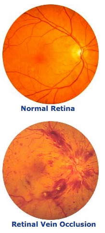
What is Retinal Vein Occlusion?
A retinal vein occlusion means that a vein in the retina of the eye has become blocked. Blockage, or occlusion, in the vein prevents adequate blood flow in the affected area. The walls of the vein leak blood and excess fluid into the retina. Central and branch retinal veins are sometimes associated with high blood pressure and diabetes.
There are two types of retinal vein occlusion:
What are the Symptoms of Retinal Vein Occlusion?
The main symptom of retinal vein occlusion is blurred vision. It occurs when the excess fluid leaking from the vein collects in the macula. The macula is the central area of the retina that is responsible for central, detailed vision. If the macula swells with excess fluid, vision blurs.
Another symptom is floaters. Floaters can appear as spots, which interfere with vision. When retinal blood vessels are not working properly, the retina may grow abnormal blood vessels (neovascularization) that are fragile. They can bleed or leak fluid into the vitreous (the gel-like fluid that fills the center of the eye), causing the floaters.
Pain in the eye sometimes occurs as a complication of severe CRVO. It is caused by excessive eye pressure called neovascular glaucoma.
How do Eye Doctors Diagnose Retinal Vein Occlusion?
After your initial examination, you may be referred to a retina specialist for further evaluation and a test of the retinal circulation called fluorescein angiography. A dye (flourescein) is injected into your arm and then special photos are taken of the inside of your eye when the dye passes through the blood vessels. Your ophthalmologist may also suggest a visit to your family physician to discover and manage any associated medical problems.
A non-invasive test called optical coherence tomography (OCT) is available at Retina Care Specialists which may help us to better evaluate how much swelling there is as a result of the branch vein occlusion.
What is the Treatment for Retinal Vein Occlusion?
In patients with macular edema from BRVO or CRVO, treatments are available that include injecting VEGF inhibitors into the eye to reduce the edema and preserve vision. The injection is painless. Sometimes, steroid injections can be used in place of or in addition to VEGF inhibitors to treat macular edema. Ozurdex is an injectable steroid implant that is FDA approved to treat macular edema.
Laser surgery improves sight in some patients with BRVO and macular edema, but vision does not usually return to normal. Laser surgery is not as helpful in CRVO.
In some cases, laser surgery is necessary to prevent vitreous hemorrhage and neovascular glaucoma. However, it does not remove hemorrhage or cure neovascular glaucoma once they are already present. It is best to treat people at risk for these complications before they occur.
Treatment is individualized based on each patient’s needs, and your physician will help you determine the best treatment available.
